
Mitochondria-Eating Protein Regulates Pathogenesis and Replication of H1N1 Influenza Virus

Updated: Jul 29
Clinical Relevance
Globally, influenza A virus (IAV) infects approximately 1 billion people every year, resulting in more than 3 million cases of severe clinical manifestations and more than 290 thousand respiratory deaths. IAV is an enveloped, negative-sense, segmented single-stranded RNA virus with a wide range of hosts including humans, birds, swine, horses, and marine mammals. The viral genome encodes 17 viral proteins including two surface glycoproteins, hemagglutinin and neuramidase, that determine viral subtype. Notably, human seasonal influenza is predominantly caused by H1N1 and H3N2 subtypes.
During IAV infection, mitochondrial dynamics are extensively altered to promote viral pathogenesis and replication. Several IAV proteins interact with mitochondrial components to induce mitochondrial fragmentation while inhibiting mitochondrial fusion. This, in turn, leads to increased reactive oxygen species (ROS) production, mitochondrial dysfunction, and subsequently cell death. ROS accumulation is required for nuclear export of viral ribonucleoprotein and viral release. Concurrently, mitophagy is activated to remove damaged mitochondria, thereby attenuating mitochondrial antiviral response including interferon signaling. Increasing evidence indicates the restoration of mitochondrial homeostasis serves as a promising therapeutic strategy for limiting viral replication and disease severity.
Potential Role of Mitochondria-Eating Protein in H1N1 Influenza Virus Infection and Preliminary Evidence
Mitochondria-eating protein (MIEAP), also known as SPATA18, is a p53-inducible protein that plays a role in mitochondrial quality control. Unlike canonical mitophagy, MIEAP promotes the accumulation of lysosomal protein and selectively repairs damaged mitochondria by degrading oxidized mitochondrial components while preserving the organelle. In addition, when the mitochondrial damage is too extensive to be repaired, MIEAP instead induces the formation of vacuoles that eliminate irreversibly damaged mitochondria, thereby maintaining mitochondrial integrity and cellular homeostasis. Although MIEAP has been characterized as a tumor suppressor in several cancer studies, its role in viral diseases remains unknown.
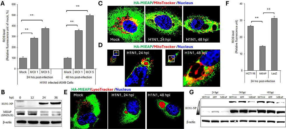
To investigate this possibility, first I confirmed that H1N1 influenza virus infection significantly increased ROS production in human lung A549 cells both in multiplicity of infection (MOI)- and time-dependent manners (Fig. A). Notably, H1N1 infection also downregulated MIEAP expression, suggesting a potential role for MIEAP in viral pathogenesis and/or replication (Fig. B). Because the commercially available SPATA18 antibody was unsuitable for confocal microscopy, I ectopically expressed an HA-tagged MIEAP (HA-MIEAP) to facilitate the visualization of its subcellular localization and dynamics. At 24 hours post-infection (24 hpi), H1N1-infected cells exhibited marked colocalization of HA-MIEAP (green) with MitoTracker Red-labeled mitochondria (Fig. C, middle panel). In addition, MIEAP formed small vacuole-like structures in the infected cell (Fig. C, middle panel). By 48 hpi, heavily infected cells developed large vacuole-like structures (Fig. C, right panel). Interestingly, mitochondrial signals were also detected within the vacuole-like structures (Fig. D). Moreover, lysotracker staining confirmed lysosomal accumulation within these vacuole-like structures, suggesting degradation of damaged cellular organelles was taking place (Fig. E). Overall, these may indicate two previously described MIEAP-associated processes: 1) mitochondrial quality control through the colocalization of MIEAP with mitochondria, indicative of a repair mechanism, and 2) the formation of MIEAP-induced vacuole (MIV), which eliminate severely damaged mitochondria.
Next, to determine whether MIEAP regulates H1N1 replication, I overexpressed MIEAP in human colorectal HCT116 cells, which exhibit little to no endogenous MIEAP expression due to promoter hypermethylation. I observed MIEAP overexpression significantly reduced H1N1-induced ROS production (Fig. F). Furthermore, compared with HA-GFP control and parental HCT116 cells, MIEAP-overexpressing cells exhibited markedly lower levels of H1N1 nucleoprotein (NP), suggesting that MIEAP suppresses H1N1 replication (Fig. G).
Take-Home Hypothesis & Clinical Implication
MIEAP is an intriguing candidate for investigation not only in H1N1 influenza but also a broad range of RNA viruses in which mitochondrial remodeling is a common feature. MIEAP represents an underexplored intersection between mitochondrial quality control, viral pathogenesis, antiviral immunity, and viral replication. One compelling hypothesis is that H1N1 suppresses MIEAP expression to preserve dysfunctional mitochondria, thereby creating a cellular environment that promotes viral replication while facilitating immune evasion. Conversely, enhancing MIEAP activity may restore mitochondrial homeostasis, limit excessive ROS production, and impair viral replication, making MIEAP a promising target for both mechanistic investigation and therapeutic intervention.
-The preliminary findings supporting this hypothesis had been presented at the Cell Symposia Mitochondria: From Signaling to Disease in Lisbon, Portugal, in 2013-
---------------------------------
HyVir would love to hear from you if this hypothesis proves to work.
Need sharper figures or references? Contact HyVir and get what you need!
Comments